AXIS Imaging Interview: Three Tips for Setting Up Remote Reading for Radiologists

Remote reading is a hot topic for many working in diagnostic imaging. It is highly desirable for many Radiologists who want to telecommute like the rest of us. And, as cloud-based imaging IT solutions become more popular, the ability to read remotely (Rads won’t be going to the data center to read) will become the standard.

AXIS Imaging interviewed me to get three tips on setting up remote reading for radiologists. The article is here.

Imaging Exam Acquisition and Quality Control (QC) Workflow in Today’s Consolidated Enterprise – Part 2 of 2

In my previous post, I discussed common challenges associated with the imaging exam acquisition workflows performed by Technologists (Tech Workflow) that many healthcare provider organizations face today.

In this post, we will explore imaging record Quality Control (QC) workflow.

Background

A typical Consolidated Enterprise is a healthcare provider organization consisting of multiple hospitals/facilities that often share a single instance of EMR/RIS and Image Manager/Archive (IM/A) systems, such as PACS or VNA. The consolidation journey is complex and requires careful planning that relies on a comprehensive approach towards a solution and interoperability architectures.

An Imaging Informatics team supporting a Consolidated Enterprise typically consists of PACS Admin and Imaging Analyst roles supporting one or more member-facilities.

Imaging Record Quality Control (QC) Workflows

To ensure the quality (completeness, consistency, correctness) of imaging records, providers rely on automatic workflows (such as validation by the IM/A system of the received DICOM study information against the corresponding HL7 patient and order information) and manual workflows performed either by Technologists during the Tech Workflow or by Imaging Informatics team members post-exam acquisition. Automatic updates of Patient and Procedure information are achieved through HL7 integration between EMR/RIS and the IM/A.

Typical manual QC activities include the following:

  • Individual Image Corrections (for example, correction of a wrong laterality marker)
  • DICOM Header Updates (for example, an update of the Study Description DICOM attribute)
  • Patient Update (moving a complete DICOM study from one patient record to another)
  • Study Merge (moving some, or all, of the DICOM objects from the “merged from” study to the “merged to” study)
  • Study Split (moving some of the DICOM objects/series from the “split from” study to the “split to” study)
  • Study Object Deletion (deletion of one or more objects/series from a study)

QC Workflow Challenges

Access Control Policy

One of the key challenges related to ensuring the quality of imaging records across large health system enterprises is determining who is qualified and authorized to perform QC activities. A common approach is to provide data control and correction tools to staff from the site where the imaging exam was acquired, since they are either aware of the context of an error or can easily get it from the interaction with the local clinical staff, systems, or the patient themselves. With such an approach, local staff can access only data acquired at sites to which they are assigned to comply with patient privacy policies and prevent any accidental updates to another site’s records. The following diagram illustrates this approach.

QC-1

Systems Responsibilities

Another important area of consideration is to determine which enterprise system should be the “source of truth” for Imaging QC workflows when there are multiple Image Manager/Archives. Consider the following common Imaging IT architecture, where multiple facilities share both PACS and VNA applications. In this scenario, the PACS maintains a local DICOM image cache while the VNA provides the long-term image archive. Both systems provide QC tools that allow authorized users to update the structure or content of imaging records.

QC-2

Since DICOM studies stored in the PACS cache also exist in the VNA, any changes resulting from QC activity performed in one of these systems must be communicated to the other to ensure that both systems are in sync. This gets more complicated when many systems storing DICOM data are involved.

Integrating the Healthcare Enterprise (IHE) developed the “Imaging Object Change Management (IOCM)” integration profile, which provides technical details regarding how to best propagate imaging record changes among multiple systems.

To minimize the complexity associated with the synchronization of imaging record changes, it is usually a good idea to appoint one system to be the “source of truth”. Although bidirectional (from PACS to VNA or from VNA to PACS) updates are technically possible, the complexity of managing and troubleshooting such integration while ensuring good data quality practices can be significant.

The Takeaway

Often the QC Workflow is not discussed in depth during the procurement phase of a new PACS or VNA. The result: The ability of the Vendor of Choice’s (VOC) solution to provide robust, reliable, and user-friendly QC tools, while ensuring compliance with access control rules across multiple sites, is not fully assessed. Practice shows that vendors vary significantly in these functional areas and their capabilities should be closely evaluated as part of any procurement process.

Imaging Exam Acquisition and Quality Control (QC) Workflow in Today’s Consolidated Enterprise – Part 1 of 2

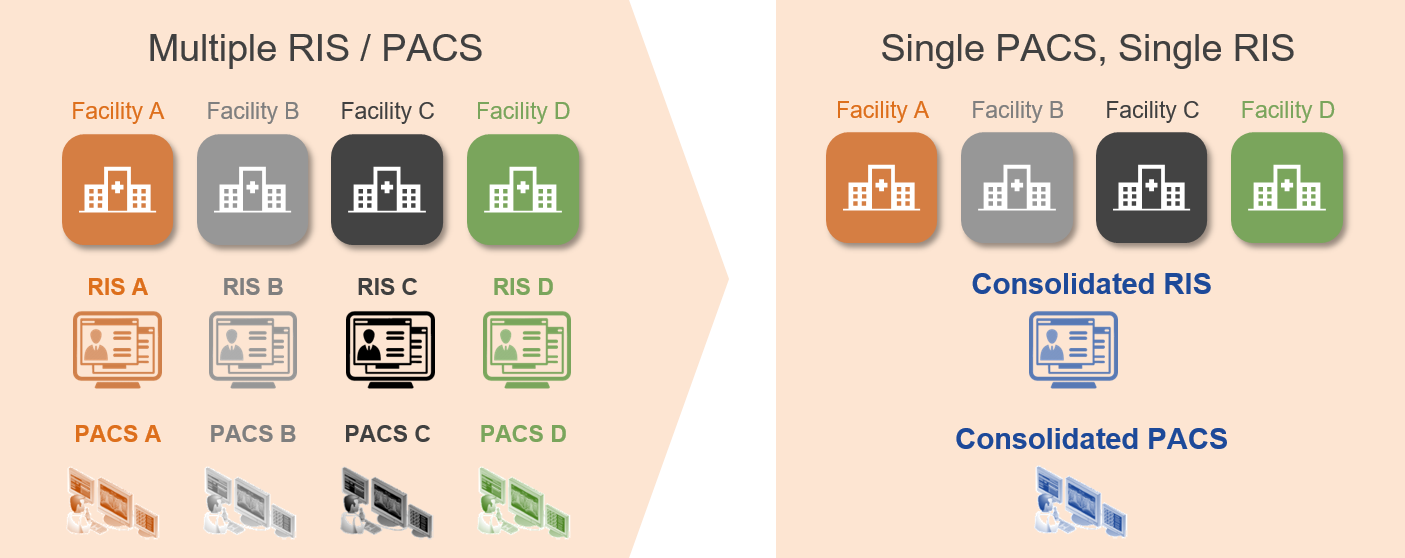
As existing healthcare provider organizations merge and affiliate to create Consolidated Enterprises, image acquisition workflows are often found to be different across the various facilities. Often, the different facilities that comprise the Consolidated Enterprise had different procedures and standard of practice for image acquisition and Quality Control (QC), along with different information and imaging systems.

Standardizing and harmonizing enterprise-wide policies, especially for imaging exam QC, can have significant benefits. A failure to standardize these workflows in a Consolidated Enterprise may result in inconsistent or inaccurate imaging records, which can lead to reading and viewing workflow challenges. These are compounded with a shared imaging system, such as an enterprise PACS or VNA, and can result in delays in care and patient safety risks.

There are generally two areas worth evaluating for optimization:

  • Technologist imaging exam acquisition workflow (Tech Workflow)
  • Imaging record Quality Control workflow (QC Workflow)

Here, we will explore Tech Workflow. QC Workflow will be covered in a subsequent post.

Throughout this discussion the term Radiology Information System (RIS) is used, which can be a standalone system or a module of an EMR.

Tech Workflow

The use of DICOM Modality Worklist (DMWL) for the management of image acquisition is well-understood and broadly adopted. However, the process of marking an exam as “complete” (or “closed”) following acquisition is less standardized and varies across different vendors and healthcare enterprises. The subsequent QC and diagnostic reading workflows rely on the “completion” of the exam before they can begin. For example, an exam that is never marked as “complete” may not appear on a Radiologist Reading Worklist, and an imaging exam that is marked as “complete” when it isn’t will be available for Radiologists to read with only a partial set of images.

Imaging Technologists typically interact with the following applications on a daily-basis.

Tech WF Screens

  • Modality Console – a comprehensive set of tools, attached to the modality, to perform image acquisition activities (such as DMWL queries, exam protocoling, post-processing, etc.).
  • Radiology Information System (RIS) – a specific view into the enterprise RIS application, allowing Technologists to look up patient/procedure information, a set of tools to document the acquisition and mark exam as “complete”, etc.
  • Image Manager/Archive (IM/A) QC – a comprehensive set of imaging exam Quality Control (QC) tools, provided by the Image Manager/Archive (IM/A), such as PACS or VNA, or a dedicated application, to make any necessary corrections to ensure the quality of acquired imaging exam records.

As stated above, there is significant variability among healthcare providers with respect to instituting Tech Workflow policies and procedures. The following diagram illustrates the steps involved in a common Tech Workflow.

Tech WF Flow

Notes:

  • In some cases, Technologists validate the quality of the image and confirm that the number of images in the IM/A is correct for multiple studies at a time instead of each one independently due to the high-volume of exams being acquired.
  • An ability to assess the quality of the imaging exam and correct it (if needed) in a quick and user-friendly manner is critical for an efficient exam completion workflow.

PACS-driven Reading Workflow

In this scenario, the PACS Client provides a Reading Worklist and it is typically responsible for launching (in-context, through a desktop integration) the Report Creator application. There are several methods used across provider organizations to communicate study complete status updates to the PACS.

Method Benefit Challenge
Time out – this is the most typical approach, which considers a study to be complete after a defined period of time has passed (for example, five minutes) since the receipt (by PACS) of the last DICOM object from the modality.
  • Easy to implement
If the time-out is too long, the creation of the corresponding Reading Worklist item will be delayed. Alternatively, a short time-out may result in a Radiologist reporting an incomplete study, which requires follow-up review and potentially an addendum to the report once the missing images are stored to PACS.
HL7 ORM – some organizations release HL7 ORM messages to the Report Creator only after the order status is updated (to study complete) in the RIS.
  • Easy to implement
  • Prevents reporting of incomplete exams (although relies on Technologists to validate the completeness of the study structure in the PACS)
There are scenarios where PACS has received DICOM studies, but their statuses in the RIS application has not yet been updated (for example, as can happen with mobile modalities). The Reading Worklist is unaware of the HL7 message flow between the RIS and the Report Creator and, therefore, allows the Radiologist to start reviewing cases. However, these cases have no corresponding procedure information in the Report Creator. When the Radiologists tries to launch the reporting application in the context of the current study, the Report Creator is unable to comply.
DICOM MPPS – Once an exam is complete, a DICOM MPPS N-Set message (issued by the modality) informs the PACS (and/or RIS) about the structure of the study and the fact that it is completed (along with other useful exam information).
  • Prevents reporting of incomplete exams
  • Automatic confirmation of the structure of the study


  • The adoption of DICOM Modality Performed Procedure Step (MPPS) is still limited in most enterprises, even though some modalities, RIS, and PACS support it.
  • Somewhat complex to implement (requires integration and testing between each modality and the MPPS server) coupled with a lack of understanding as to the benefits of this approach in many healthcare provider organizations.
  • Some modality vendors charge an additional fee for a license to enable MPPS integration.
  • Can be disconnected from the “completion” of the exam in RIS (i.e. can ensure the Report Creator’s readiness), provided only the PACS receives and processes the MPPS messages.
DICOM Storage Commitment – Once the exam is complete, a series of DICOM messages (N-Action, N-Event-Report) between modalities and PACS can determine whether a complete study was stored to PACS.
  • Prevents reporting of incomplete exams
  • Automatic confirmation of the structure of the study
  • Although most PACS and many modalities support this DICOM transaction, it is not widely implemented by healthcare providers.
  • Somewhat complex to implement (requires integration and testing between each modality and the PACS server) coupled with a lack of understanding as to the benefits of this approach in many healthcare provider organizations.
  • Can be disconnected from the “completion” of the exam in RIS (i.e. can ensure the Report Creator’s readiness).

RIS-driven Reading Workflow

In this scenario, the RIS provides the Reading Worklist and it is implicitly aware of the status of the exam (assuming the same system is used by Techs and Rads). It creates the worklist item that corresponds to the exam once it reaches the “complete” status. As the Reading Worklist launches both the Report Creator and the Diagnostic Viewer (PACS Client) applications, it does not face the informatics challenges inherent to the PACS-driven Reading Workflow described above.

Enterprise-wide Reading Workflow (Dedicated, Standalone Application)

Some organizations use an enterprise-wide Reading Worklist that is a separate application from the PACS and RIS to orchestrate enterprise-wide diagnostic reading (and other imaging related) tasks across all their Radiologists using fine-grained task-allocation rules. Similar to the RIS-driven Reading Workflow, the worklist launches both the Report Creator and the Diagnostic Viewer applications once a worklist item is selected.

To prevent the complexity of the PACS-driven Reading Workflow described above, some organizations choose to release an HL7 ORM message from the RIS application to the worklist only when the status of the corresponding exam in that system is updated. Alternatively, organizations that choose to send all ORM messages to the worklist application as soon as procedures are scheduled, need to deal with ensuring that the PACS has a complete study prior to allowing it to be reported.

So, what?

It is important for healthcare provider organizations to understand the relationship between the Tech Workflow and the Reading Worklist approach they adopt. If a RIS-driven approach is not chosen, then there should be a clear integration strategy in place to ensure that studies are not reported too soon or missed.

SIIM 2017 – Economics of Imaging Informatics

I feel fortunate to chair a very interesting and relevant session during the SIIM 2017 annual meeting this week. The session will focus on the following areas, which are often discussed with clients and industry colleagues.

It seems that there is no well-defined formula or consensus regarding the staffing levels and the structure of an Imaging Informatics Team (IIT). Typical questions are:

  • How large should an IIT be, given the annual Radiology exam volume and number facilities the team supports?
  • In the era of EMRs and IT centralization, what is the right mix of roles between IIT and Corporate IT?
  • How does IT centralization affect IIT financial decision-making and budgets?

James Forrester’s talk will address the above topics, based on his experience at University of Rochester Medical Center.

Another interesting observation is that typically outside of the Director of IIT role, IIT members lack understanding of corporate finance. Even though most significant projects must be budgeted for in advance and any innovation or improvement proposal must be accompanied with a well-crafted business case. Robert (Bob) Coleman’s talk will provide an overview of proven budgeting methods, core components of a good business case, and the required skills to prepare one.

You may also wish to check out this interactive, whiteboard session, which will walk through some common financial models used to evaluate the Total Cost of Ownership (TCO) and Return on Investment (ROI) for imaging IT projects when talking to the CIO.

Looking forward to seeing everybody in Pittsburgh!

Dealing with Multiple Terminology Domains in a Consolidated Enterprise – Part 2

In my previous post, Dealing with Multiple Terminology Domains in a Consolidated Enterprise, I introduced a typical challenge that many imaging projects face today.

In this post, I will describe three common use cases where the problem of multiple terminology domains manifests.

Single PACS, Multiple RIS

Often, rapidly growing health systems aim to consolidate imaging informatics solutions across their facilities. Replacement of multiple PACS with one such system, while keeping separate RIS systems in place is not uncommon. The reason behind this dichotomy is that a RIS is much more ingrained into the local Radiology department’s operational and clinical workflows than a PACS, making its replacement complex and impactful on many stakeholders.

The following diagram illustrates this scenario.

term-pacs

In such a deployment, the consolidated PACS is responsible for dealing with multiple ordering systems that use individual procedure terminologies. It also maintains patients’ longitudinal imaging record, which will include different values in the DICOM headers to describe the same procedure types.

Multiple RIS/PACS, Shared VNA

Health systems that seek to benefit from IT infrastructure consolidation, as well as a single Imaging Record Management, Archive, Access, and Sharing application, often opt to procure and deploy a shared VNA system across their facilities. By keeping their RIS/PACS systems in place they can rapidly deliver clinical and operational benefits with minimal disruption to the existing workflows. This approach allows individual facilities to stay fairly independent in their imaging informatics system and process decision making.

The following diagram illustrates this scenario.

term-vna

In this deployment, the shared VNA typically maps or normalizes procedure terminologies in the DICOM header of the studies that are served to the individual PACS systems as part of the relevant prior pre-/push-fetch workflows.

Single PACS, Single RIS

An increasingly common scenario is when health systems include a RIS consolidation project within their EMR consolidation strategy, while PACS consolidation happens in parallel. This approach results in a single master set of orderable procedures that is used by all participating facilities. The challenge arises from the fact that historic imaging records maintain, in the DICOM data, procedure information using historic terminology values that predate consolidation and can include known values (from the latest RIS) or some potentially unknown value (previous RIS systems for the institutions that replaced their RIS system at least once and did not replace the values with one used by the new RIS).

The following diagram illustrates this scenario.

term-rispng

In these deployments, the consolidated PACS is responsible for dealing with new common and fragmented historic procedure terminologies.

In the next post, I will describe how PACS and VNA vendors deal with this challenge.

RSNA 2016

rsna_2016_logo_dates_rgb

In what I believe is my 15th consecutive RSNA, I have a full schedule of business meetings, committee and board meetings, with some time for connecting with friends. In addition to the typical, semi-organized chaos, I am giving two talks.

Hope to see you all in Chicago.

RCC24 – Starting a Health IT Consulting Company

Room: S501ABC
Mon 28-Nov-2016, 2:30 pm to 4:00 pm CT

RC654 – Using IHE Profiles to Plan for Medical Imaging

Room: S504AB
Thu 1-Dec-2016, 8:30 am to 10:00 am CT
chicago-rsna

Dealing with Multiple Terminology Domains in a Consolidated Enterprise

As the number of the PACS consolidation projects grow, I think it is important to explore some of the informatics concepts that need to be addressed to maximize the value of a consolidated PACS’ clinical functionality.

As mentioned in my recent MIIT talk, there are operational, financial and clinical goals that drive PACS consolidation projects. One of those reasons is to enable multi-facility diagnostic reading workflow: acquire anywhere and read anywhere in the enterprise.

One of the key informatics prerequisites of a successful PACS consolidation project is dealing with Patient Identities in a Consolidated Enterprise to establish patients’ longitudinal imaging record. Once that fundamental challenge is addressed, dealing with the normalization or mapping of the exam terminologies used by different RIS systems across the consolidated enterprise is the next critical informatics area to tackle. Often, PACS consolidation projects do not include the unification of the facility RIS, which forces the PACS to deal with multiple terminology domains.

In this series of the blog posts, I will examine this challenge in detail and describe the imaging informatics industry’s current capabilities to deal with it.

The Challenge

First of all, let’s define the problem and why it is important.

The anatomical and procedural information for a radiology exam is used by the PACS to primarily: 1) determine relevancy across patients’ historic studies; and 2) establish the correct display protocol for the PACS Workstation. As different ordering systems (EMR/RIS) may use different values to describe the same ordered procedure, the consolidated PACS will have to use a value normalization or mapping method to properly process the information.

The following diagram conceptually illustrates the difference between normalization and mapping methods.

terminology

Mapping

This approach relies on keeping many-to-many translation tables where each term has a corresponding defined value under each terminology domain. This approach is feasible only with a very small number of values and terminology domains.

Normalization

This methodology creates a “canonical” representation of each term and establishes a one-to-one relationship between each value in each terminology domain and the corresponding value under the “canonical” representation. This approach can accommodate a very large number of values and terminologies, as the translation from one terminology to another is always done through the canonical value.

In the next post, I will describe the imaging informatics use-cases that have to deal with this challenge.

Article: Major Insurance Company will no longer cover Breast Tomosynthesis Exams

Per this article, citing lack of clinical evidence and radiation dose concerns, Cigna will no longer cover Breast Tomosynthesis exams, which generate 3D image sets, as of 15-Feb-2016.

Digital Breast Tomosynthesis (DBT) exams are often praised for their superior ability to provide effective imaging when diagnosing women with dense breasts.

Somewhat of a challenge for IT systems and staff due to the significant size of their data sets compared to the traditional 2D mammogram exams, adoption of DBT modalities has been rapid lately due to the generally accepted diagnostic benefits.

I can’t imagine that this lack of coverage will last.

For those wanting a description of the different between 2D (Mammogram) and 3D (Tomosynthesis) breast imaging, this article provides an overview in plain language.

Here is an RSNA article on the state-of-the-art for Breast Tomosynthesis.

And here is an article on the merits of Breast Tomosynthesis over traditional Mammography for detecting cancer.

And another on reducing recall rates.