Imaging Exam Acquisition and Quality Control (QC) Workflow in Today’s Consolidated Enterprise – Part 2 of 2

In my previous post, I discussed common challenges associated with the imaging exam acquisition workflows performed by Technologists (Tech Workflow) that many healthcare provider organizations face today.

In this post, we will explore imaging record Quality Control (QC) workflow.

Background

A typical Consolidated Enterprise is a healthcare provider organization consisting of multiple hospitals/facilities that often share a single instance of EMR/RIS and Image Manager/Archive (IM/A) systems, such as PACS or VNA. The consolidation journey is complex and requires careful planning that relies on a comprehensive approach towards a solution and interoperability architectures.

An Imaging Informatics team supporting a Consolidated Enterprise typically consists of PACS Admin and Imaging Analyst roles supporting one or more member-facilities.

Imaging Record Quality Control (QC) Workflows

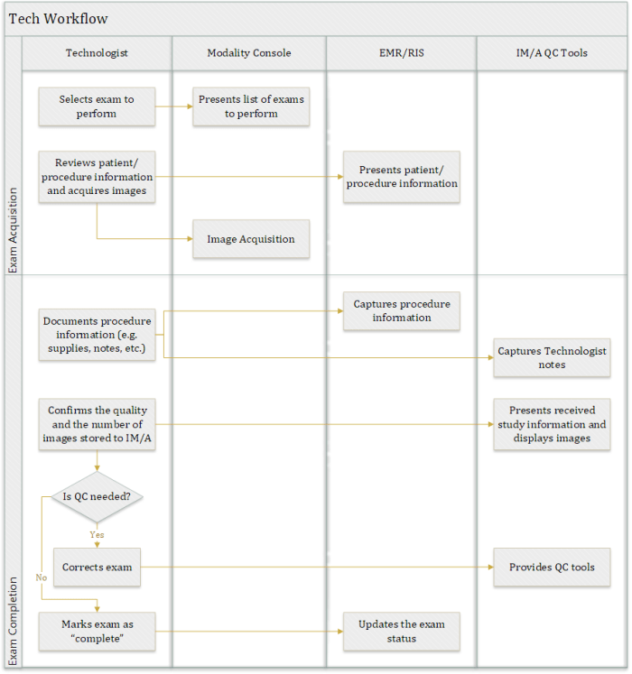
To ensure the quality (completeness, consistency, correctness) of imaging records, providers rely on automatic workflows (such as validation by the IM/A system of the received DICOM study information against the corresponding HL7 patient and order information) and manual workflows performed either by Technologists during the Tech Workflow or by Imaging Informatics team members post-exam acquisition. Automatic updates of Patient and Procedure information are achieved through HL7 integration between EMR/RIS and the IM/A.

Typical manual QC activities include the following:

  • Individual Image Corrections (for example, correction of a wrong laterality marker)
  • DICOM Header Updates (for example, an update of the Study Description DICOM attribute)
  • Patient Update (moving a complete DICOM study from one patient record to another)
  • Study Merge (moving some, or all, of the DICOM objects from the “merged from” study to the “merged to” study)
  • Study Split (moving some of the DICOM objects/series from the “split from” study to the “split to” study)
  • Study Object Deletion (deletion of one or more objects/series from a study)

QC Workflow Challenges

Access Control Policy

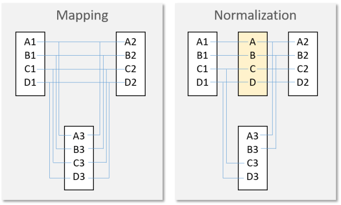
One of the key challenges related to ensuring the quality of imaging records across large health system enterprises is determining who is qualified and authorized to perform QC activities. A common approach is to provide data control and correction tools to staff from the site where the imaging exam was acquired, since they are either aware of the context of an error or can easily get it from the interaction with the local clinical staff, systems, or the patient themselves. With such an approach, local staff can access only data acquired at sites to which they are assigned to comply with patient privacy policies and prevent any accidental updates to another site’s records. The following diagram illustrates this approach.

QC-1

Systems Responsibilities

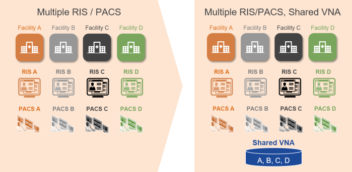
Another important area of consideration is to determine which enterprise system should be the “source of truth” for Imaging QC workflows when there are multiple Image Manager/Archives. Consider the following common Imaging IT architecture, where multiple facilities share both PACS and VNA applications. In this scenario, the PACS maintains a local DICOM image cache while the VNA provides the long-term image archive. Both systems provide QC tools that allow authorized users to update the structure or content of imaging records.

QC-2

Since DICOM studies stored in the PACS cache also exist in the VNA, any changes resulting from QC activity performed in one of these systems must be communicated to the other to ensure that both systems are in sync. This gets more complicated when many systems storing DICOM data are involved.

Integrating the Healthcare Enterprise (IHE) developed the “Imaging Object Change Management (IOCM)” integration profile, which provides technical details regarding how to best propagate imaging record changes among multiple systems.

To minimize the complexity associated with the synchronization of imaging record changes, it is usually a good idea to appoint one system to be the “source of truth”. Although bidirectional (from PACS to VNA or from VNA to PACS) updates are technically possible, the complexity of managing and troubleshooting such integration while ensuring good data quality practices can be significant.

The Takeaway

Often the QC Workflow is not discussed in depth during the procurement phase of a new PACS or VNA. The result: The ability of the Vendor of Choice’s (VOC) solution to provide robust, reliable, and user-friendly QC tools, while ensuring compliance with access control rules across multiple sites, is not fully assessed. Practice shows that vendors vary significantly in these functional areas and their capabilities should be closely evaluated as part of any procurement process.

Imaging Exam Acquisition and Quality Control (QC) Workflow in Today’s Consolidated Enterprise – Part 1 of 2

As existing healthcare provider organizations merge and affiliate to create Consolidated Enterprises, image acquisition workflows are often found to be different across the various facilities. Often, the different facilities that comprise the Consolidated Enterprise had different procedures and standard of practice for image acquisition and Quality Control (QC), along with different information and imaging systems.

Standardizing and harmonizing enterprise-wide policies, especially for imaging exam QC, can have significant benefits. A failure to standardize these workflows in a Consolidated Enterprise may result in inconsistent or inaccurate imaging records, which can lead to reading and viewing workflow challenges. These are compounded with a shared imaging system, such as an enterprise PACS or VNA, and can result in delays in care and patient safety risks.

There are generally two areas worth evaluating for optimization:

  • Technologist imaging exam acquisition workflow (Tech Workflow)
  • Imaging record Quality Control workflow (QC Workflow)

Here, we will explore Tech Workflow. QC Workflow will be covered in a subsequent post.

Throughout this discussion the term Radiology Information System (RIS) is used, which can be a standalone system or a module of an EMR.

Tech Workflow

The use of DICOM Modality Worklist (DMWL) for the management of image acquisition is well-understood and broadly adopted. However, the process of marking an exam as “complete” (or “closed”) following acquisition is less standardized and varies across different vendors and healthcare enterprises. The subsequent QC and diagnostic reading workflows rely on the “completion” of the exam before they can begin. For example, an exam that is never marked as “complete” may not appear on a Radiologist Reading Worklist, and an imaging exam that is marked as “complete” when it isn’t will be available for Radiologists to read with only a partial set of images.

Imaging Technologists typically interact with the following applications on a daily-basis.

Tech WF Screens

  • Modality Console – a comprehensive set of tools, attached to the modality, to perform image acquisition activities (such as DMWL queries, exam protocoling, post-processing, etc.).
  • Radiology Information System (RIS) – a specific view into the enterprise RIS application, allowing Technologists to look up patient/procedure information, a set of tools to document the acquisition and mark exam as “complete”, etc.
  • Image Manager/Archive (IM/A) QC – a comprehensive set of imaging exam Quality Control (QC) tools, provided by the Image Manager/Archive (IM/A), such as PACS or VNA, or a dedicated application, to make any necessary corrections to ensure the quality of acquired imaging exam records.

As stated above, there is significant variability among healthcare providers with respect to instituting Tech Workflow policies and procedures. The following diagram illustrates the steps involved in a common Tech Workflow.

Tech WF Flow

Notes:

  • In some cases, Technologists validate the quality of the image and confirm that the number of images in the IM/A is correct for multiple studies at a time instead of each one independently due to the high-volume of exams being acquired.
  • An ability to assess the quality of the imaging exam and correct it (if needed) in a quick and user-friendly manner is critical for an efficient exam completion workflow.

PACS-driven Reading Workflow

In this scenario, the PACS Client provides a Reading Worklist and it is typically responsible for launching (in-context, through a desktop integration) the Report Creator application. There are several methods used across provider organizations to communicate study complete status updates to the PACS.

Method Benefit Challenge
Time out – this is the most typical approach, which considers a study to be complete after a defined period of time has passed (for example, five minutes) since the receipt (by PACS) of the last DICOM object from the modality.
  • Easy to implement
If the time-out is too long, the creation of the corresponding Reading Worklist item will be delayed. Alternatively, a short time-out may result in a Radiologist reporting an incomplete study, which requires follow-up review and potentially an addendum to the report once the missing images are stored to PACS.
HL7 ORM – some organizations release HL7 ORM messages to the Report Creator only after the order status is updated (to study complete) in the RIS.
  • Easy to implement
  • Prevents reporting of incomplete exams (although relies on Technologists to validate the completeness of the study structure in the PACS)
There are scenarios where PACS has received DICOM studies, but their statuses in the RIS application has not yet been updated (for example, as can happen with mobile modalities). The Reading Worklist is unaware of the HL7 message flow between the RIS and the Report Creator and, therefore, allows the Radiologist to start reviewing cases. However, these cases have no corresponding procedure information in the Report Creator. When the Radiologists tries to launch the reporting application in the context of the current study, the Report Creator is unable to comply.
DICOM MPPS – Once an exam is complete, a DICOM MPPS N-Set message (issued by the modality) informs the PACS (and/or RIS) about the structure of the study and the fact that it is completed (along with other useful exam information).
  • Prevents reporting of incomplete exams
  • Automatic confirmation of the structure of the study


  • The adoption of DICOM Modality Performed Procedure Step (MPPS) is still limited in most enterprises, even though some modalities, RIS, and PACS support it.
  • Somewhat complex to implement (requires integration and testing between each modality and the MPPS server) coupled with a lack of understanding as to the benefits of this approach in many healthcare provider organizations.
  • Some modality vendors charge an additional fee for a license to enable MPPS integration.
  • Can be disconnected from the “completion” of the exam in RIS (i.e. can ensure the Report Creator’s readiness), provided only the PACS receives and processes the MPPS messages.
DICOM Storage Commitment – Once the exam is complete, a series of DICOM messages (N-Action, N-Event-Report) between modalities and PACS can determine whether a complete study was stored to PACS.
  • Prevents reporting of incomplete exams
  • Automatic confirmation of the structure of the study
  • Although most PACS and many modalities support this DICOM transaction, it is not widely implemented by healthcare providers.
  • Somewhat complex to implement (requires integration and testing between each modality and the PACS server) coupled with a lack of understanding as to the benefits of this approach in many healthcare provider organizations.
  • Can be disconnected from the “completion” of the exam in RIS (i.e. can ensure the Report Creator’s readiness).

RIS-driven Reading Workflow

In this scenario, the RIS provides the Reading Worklist and it is implicitly aware of the status of the exam (assuming the same system is used by Techs and Rads). It creates the worklist item that corresponds to the exam once it reaches the “complete” status. As the Reading Worklist launches both the Report Creator and the Diagnostic Viewer (PACS Client) applications, it does not face the informatics challenges inherent to the PACS-driven Reading Workflow described above.

Enterprise-wide Reading Workflow (Dedicated, Standalone Application)

Some organizations use an enterprise-wide Reading Worklist that is a separate application from the PACS and RIS to orchestrate enterprise-wide diagnostic reading (and other imaging related) tasks across all their Radiologists using fine-grained task-allocation rules. Similar to the RIS-driven Reading Workflow, the worklist launches both the Report Creator and the Diagnostic Viewer applications once a worklist item is selected.

To prevent the complexity of the PACS-driven Reading Workflow described above, some organizations choose to release an HL7 ORM message from the RIS application to the worklist only when the status of the corresponding exam in that system is updated. Alternatively, organizations that choose to send all ORM messages to the worklist application as soon as procedures are scheduled, need to deal with ensuring that the PACS has a complete study prior to allowing it to be reported.

So, what?

It is important for healthcare provider organizations to understand the relationship between the Tech Workflow and the Reading Worklist approach they adopt. If a RIS-driven approach is not chosen, then there should be a clear integration strategy in place to ensure that studies are not reported too soon or missed.

SIIM 2017 – Economics of Imaging Informatics

I feel fortunate to chair a very interesting and relevant session during the SIIM 2017 annual meeting this week. The session will focus on the following areas, which are often discussed with clients and industry colleagues.

It seems that there is no well-defined formula or consensus regarding the staffing levels and the structure of an Imaging Informatics Team (IIT). Typical questions are:

  • How large should an IIT be, given the annual Radiology exam volume and number facilities the team supports?
  • In the era of EMRs and IT centralization, what is the right mix of roles between IIT and Corporate IT?
  • How does IT centralization affect IIT financial decision-making and budgets?

James Forrester’s talk will address the above topics, based on his experience at University of Rochester Medical Center.

Another interesting observation is that typically outside of the Director of IIT role, IIT members lack understanding of corporate finance. Even though most significant projects must be budgeted for in advance and any innovation or improvement proposal must be accompanied with a well-crafted business case. Robert (Bob) Coleman’s talk will provide an overview of proven budgeting methods, core components of a good business case, and the required skills to prepare one.

You may also wish to check out this interactive, whiteboard session, which will walk through some common financial models used to evaluate the Total Cost of Ownership (TCO) and Return on Investment (ROI) for imaging IT projects when talking to the CIO.

Looking forward to seeing everybody in Pittsburgh!

Dealing with Multiple Terminology Domains in a Consolidated Enterprise – Part 2

In my previous post, Dealing with Multiple Terminology Domains in a Consolidated Enterprise, I introduced a typical challenge that many imaging projects face today.

In this post, I will describe three common use cases where the problem of multiple terminology domains manifests.

Single PACS, Multiple RIS

Often, rapidly growing health systems aim to consolidate imaging informatics solutions across their facilities. Replacement of multiple PACS with one such system, while keeping separate RIS systems in place is not uncommon. The reason behind this dichotomy is that a RIS is much more ingrained into the local Radiology department’s operational and clinical workflows than a PACS, making its replacement complex and impactful on many stakeholders.

The following diagram illustrates this scenario.

term-pacs

In such a deployment, the consolidated PACS is responsible for dealing with multiple ordering systems that use individual procedure terminologies. It also maintains patients’ longitudinal imaging record, which will include different values in the DICOM headers to describe the same procedure types.

Multiple RIS/PACS, Shared VNA

Health systems that seek to benefit from IT infrastructure consolidation, as well as a single Imaging Record Management, Archive, Access, and Sharing application, often opt to procure and deploy a shared VNA system across their facilities. By keeping their RIS/PACS systems in place they can rapidly deliver clinical and operational benefits with minimal disruption to the existing workflows. This approach allows individual facilities to stay fairly independent in their imaging informatics system and process decision making.

The following diagram illustrates this scenario.

term-vna

In this deployment, the shared VNA typically maps or normalizes procedure terminologies in the DICOM header of the studies that are served to the individual PACS systems as part of the relevant prior pre-/push-fetch workflows.

Single PACS, Single RIS

An increasingly common scenario is when health systems include a RIS consolidation project within their EMR consolidation strategy, while PACS consolidation happens in parallel. This approach results in a single master set of orderable procedures that is used by all participating facilities. The challenge arises from the fact that historic imaging records maintain, in the DICOM data, procedure information using historic terminology values that predate consolidation and can include known values (from the latest RIS) or some potentially unknown value (previous RIS systems for the institutions that replaced their RIS system at least once and did not replace the values with one used by the new RIS).

The following diagram illustrates this scenario.

term-rispng

In these deployments, the consolidated PACS is responsible for dealing with new common and fragmented historic procedure terminologies.

In the next post, I will describe how PACS and VNA vendors deal with this challenge.

Dealing with Multiple Terminology Domains in a Consolidated Enterprise

As the number of the PACS consolidation projects grow, I think it is important to explore some of the informatics concepts that need to be addressed to maximize the value of a consolidated PACS’ clinical functionality.

As mentioned in my recent MIIT talk, there are operational, financial and clinical goals that drive PACS consolidation projects. One of those reasons is to enable multi-facility diagnostic reading workflow: acquire anywhere and read anywhere in the enterprise.

One of the key informatics prerequisites of a successful PACS consolidation project is dealing with Patient Identities in a Consolidated Enterprise to establish patients’ longitudinal imaging record. Once that fundamental challenge is addressed, dealing with the normalization or mapping of the exam terminologies used by different RIS systems across the consolidated enterprise is the next critical informatics area to tackle. Often, PACS consolidation projects do not include the unification of the facility RIS, which forces the PACS to deal with multiple terminology domains.

In this series of the blog posts, I will examine this challenge in detail and describe the imaging informatics industry’s current capabilities to deal with it.

The Challenge

First of all, let’s define the problem and why it is important.

The anatomical and procedural information for a radiology exam is used by the PACS to primarily: 1) determine relevancy across patients’ historic studies; and 2) establish the correct display protocol for the PACS Workstation. As different ordering systems (EMR/RIS) may use different values to describe the same ordered procedure, the consolidated PACS will have to use a value normalization or mapping method to properly process the information.

The following diagram conceptually illustrates the difference between normalization and mapping methods.

terminology

Mapping

This approach relies on keeping many-to-many translation tables where each term has a corresponding defined value under each terminology domain. This approach is feasible only with a very small number of values and terminology domains.

Normalization

This methodology creates a “canonical” representation of each term and establishes a one-to-one relationship between each value in each terminology domain and the corresponding value under the “canonical” representation. This approach can accommodate a very large number of values and terminologies, as the translation from one terminology to another is always done through the canonical value.

In the next post, I will describe the imaging informatics use-cases that have to deal with this challenge.

VNA and Enterprise Viewer Projects’ ROI

When I discuss industry trends with colleagues and clients, I find that we periodically touch on the topic of defining and realizing VNA and Enterprise Viewer (EV) projects’ return on investment (ROI). Our industry has made several attempts to develop an ROI calculator, which would typically encompass:

  • the benefits of consolidating IT infrastructure;
  • avoiding the cost of repeat exams due to the availability of a longitudinal patient imaging record;
  • and efficiency gains stemming from the optimized distribution and visualization of medical images.

Often these calculations are tied to a specific project and not easily reused.

During our involvement in various VNA and EV projects, we observed an interesting pattern that can bring an additional perspective on the ROI discussion.

By the end of 2010, the vast majority of U.S. hospitals had installed a PACS solution. The bulk of the deployments took place during the 2005-2010 period, and many of those are still in place, bolstered by many upgrades and technology-refresh cycles since their initial installations. During that period, both the hardware and storage components of a PACS solution were often procured directly from the PACS vendors. This procurement approach allowed the vendors to enjoy significant Service and Maintenance Agreement (SMA) revenues that would cover not only their solution components but also any included third-party hardware and storage.

Since that procurement wave, many things have changed:

  • PACS market maturity resulted in a commoditization of some of its functional areas
  • Hardware and storage costs have significantly dropped
  • Server virtualization became the preferred deployment methodology
  • Procurement of the infrastructure components has been steadily shifting from the Radiology department to the Enterprise/Corporate IT team

Also, PACS market saturation depreciated PACS vendors’ software license sales, resulting in SMA revenues becoming the key contributor to their top line.

All of these changes often created a tension between a hospital’s staff and its PACS vendor because the perceived value of the services delivered under the SMA contracts do not seem to warrant the high dollar cost. Besides tough negotiation tactics, a hospital has few practical tools at its disposal to change this dynamic. This is where well-thought-out VNA and EV projects may become extremely important in changing the negotiation power balance.

The technical and operational benefits of having a VNA take over a PACS Archive, EMR integration and sometimes even workflow components by the VNA and EV solutions are well documented and often result in the hospital’s reduced dependency on the existing PACS vendor.

Consequently, a hospital that implements VNA and EV solutions will be well-suited to renegotiate existing PACS SMA contracts to adequately reflect the provided service. The reduced SMA value can partially offset the cost of VNA and EV projects, thus contributing positively to the ROI calculation. Having said that, without a compelling event, such as an RFP to replace the existing PACS, the incumbent vendor will have little incentive to concede in the SMA renegotiations.

In order to successfully realize the above potential savings, it is important to understand what core functional areas of a PACS can be replaced by a VNA or an EV solution. Consider the following diagram:

ROI-1

Impact on Workflow or External Systems Replacement Complexity Industry Ability to Replace
Long-term Archiving and ILM This functionality is typically not exposed to external systems and has relatively simple orchestration workflows Low: Besides the need to keep the VNA copy of the study in sync with the one cached by the PACS, the archival and retrieval functionality is relatively straight-forward Current state-of-the-art VNA solutions offer proven methodologies to take over this functional area from the PACS
Routing, Pre-fetching and Relevancy This functional area may play an important role in orchestrating a departmental or an enterprise workflow Moderate: Relevancy detection can potentially increase the relative complexity of study routing and pre-fetching, which are typically quite straightforward due to their transactional nature The majority of the leading VNA solutions can adequately deliver this functionality, but their rule-definition flexibility coupled with their ability to express sophisticated relevancy rules (especially across multiple terminology domains), may vary
Acquisition and Quality Control (QC) Workflow Orchestration This functionality has a major impact on the acquisition and reading workflow with a large number of 3rd party systems integrations High: The large number of acquisition modalities will often have different associated configurations. Additionally, in large enterprises QC workflows could be very complex involving both automatic and manual activities. The effort to recreate all QC workflows, which were accumulated over the course of many years could be quite significant The VNA systems’ ability to provide this functionality represents one of the major product differentiation areas among current vendors
Image Distribution and EMR Integration An ability to provide access to images outside of the Radiology department is a critical component of a provider’s single patient record objective Low: The need to provide access to images within multiple applications (e.g. EMR, portal) or stand-alone impose some security and integration challenges. Besides the privacy and security considerations, the rest of the deployment and integration activities are relatively straight forward. Current state-of-the-art EV solutions offer proven methodologies to take over this functional area from PACS

Although this post is primarily focused on SMA-related costs, the reduction of the PACS functional scope will also decrease the corresponding Professional Services expenses.

Working on an Enterprise Imaging project? Leave us a comment with your thoughts, or contact us.

Developing an Enterprise Imaging Strategy—What is the best approach?

In my last post, we explored the current state-of-the-art of the Enterprise Imaging (EI) industry. In this post, I will zoom in on storing and managing non-DICOM images and videos. This can be ambiguous and may confuse providers who are trying to procure an EI solution. It also results in different schools of thought among vendors.

Currently, EI content can be stored and managed in one of the following formats:

  • Original object (e.g. jpg) stored in a solution’s database and/or filesystem using the vendor’s API (Application Programming Interface)
  • Original object (e.g. jpg) stored using the IHE Cross-Enterprise Document Sharing (XDS) integration profile in a solution’s XDS Document Repository component
  • Original object (e.g. jpg) stored in a solution’s database and/or filesystem using HL7’s FHIR® Media Content specifications
  • DICOM object stored in a solution’s Image Manager/Archive component; for example, using the IHE Web Image Capture (WIC) integration profile
  • DICOM object stored in a solution’s Image Manager/Archive and XDS Document Repository components using the IHE Cross-Enterprise Document Sharing for Imaging (XDS-I) integration profile

The following diagram depicts the main steps that take place during information capture activity for each method.

storage methods

All of the above methods have corresponding pros and cons, which leads to the current divergence of opinions regarding the best option to use. Having said that, it is clear that, irrespective of the chosen method, there is a need to properly collect and manage patient, administrative and clinical context (aka metadata) for the acquired EI content.

Metadata

Each of the above methods offer different levels of metadata rigidity and extensibility which impact the interoperability:

  • DICOM, FHIR and XDS-I based methods offer a level of certainty for the vendors with respect to what information should be captured and how it should be encoded.
  • XDS takes an approach of developing specific content profiles that address specific types of content; for example, the IHE XDS-SD (Scanned Document) integration profile. At the moment, there is no content profile that is specific to the Enterprise Imaging domain. Additionally, XDS allows the original object to be wrapped in a CDA Document to capture additional metadata in case the specified XDS Document Entry attributes are not sufficient.

Is there one “right” answer?

There are two overarching clinical reasons to capture EI content:

  1. To enrich patients’ clinical record
  2. To provide reliable, authorized access to it across the enterprise (and beyond)

As the following diagram suggests, the way EI content is stored is less important then the flexibility of an EI solution’s “Capture” and “Discovery and Access” components, because it is hidden behind those interfaces.

EI Access

It seems that, currently, there is no single answer for the best EI content format given the informatics complexity of healthcare provider’s enterprises. In order to adapt and compete, vendors will be pressured to support multiple inbound and outbound methods (such as FHIR, DICOM, DICOMWeb, XDS, proprietary APIs, etc.) and only time will tell which approach will become a de-facto standard.

Working on an Enterprise Imaging project? Leave us a comment with your thoughts, or contact us.

Enterprise Imaging Industry State-of-the-Art

Based on discussions with colleagues and our clients, Enterprise Imaging is becoming an integral part of U.S. Hospital IT Consolidated Clinical Record strategies.

HIMSS-SIIM Enterprise Imaging Workgroup‘s current working definition of Enterprise Imaging is as following:

  • Diagnostic Imaging – Encompassing traditional diagnostic imaging disciplines such as Radiology and Cardiology
  • Procedural Imaging – Including images that are acquired for diagnosis or clinical documentation purposes (such as visible light photos, point-of-care ultrasound)
  • Evidence Imaging – Including images and/or videos that are acquired for clinical documentation purposes (for example, scope videos, computer aided detection)
  • Image-based Clinical Reports – Documentation that includes or entirely consists of images (for example, Pulmonary Functional Test (PFT) report, multi-media pathology report)

Despite the attention from vendors, industry focus, and provider demand, this market space is still in its early stages of development. There are two main reasons: 1) the scope of the problem domain is still being defined; and 2) the vendor community is still working out the best practices and optimal technical approaches.

Moreover, the number of the departments that generate Enterprise Imaging content and that have their own departmental workflows is quite large.

This results in significant confusion on the provider side who are left to navigate a myriad of perspectives expressed by the imaging informatics industry. There are on-going initiatives that are currently working on demystifying the field of Enterprise Imaging. For example, the recent SIIM Webinar delivered by Dr. Towbin from Cincinnati Children’s, provides a very thorough analysis of the problem domain.

In conversations with vendors and providers, we have compiled several observations that might benefit the imaging informatics community.

The Right Approach

In the SIIM 2015 Opening General Session presentation, Don Dennison presented the following slide titled “Enterprise Image Management: Making the Right Choice”

EI

With the various systems in place to manage patient record data, there is often debate as to which enterprise system is best suited to offer Enterprise Imaging services.

At the moment, there is no obvious answer to the question presented by the slide. Besides the technical capabilities of the systems, the provider’s internal IT capabilities, capacity and policies can significantly influence the decision. At some organizations, where the Imaging Informatics Team plays a prominent IT role, the choice could be the VNA, while at others, where the Enterprise IT team takes the lead, the EMR or ECM is often chosen.

The Right Functionality

During RSNA 2015, we conducted a study to identify the state-of-the-art of Enterprise Imaging technology, including methods of acquisition, management, and distribution of non-DICOM images. The following table summarizes our findings.


Image / Video Acquisition
Ability to capture from mobile devices The majority of current vendors opted for native applications to provide better user experience and tighter security controls. Still, image capture is the prevailing capability, with video acquisition capabilities lagging behind. Some vendors offer integration with leading EMRs’ mobile applications.
Ability to capture from visible light cameras The ability to manually (i.e. file browse, drag & drop, etc.) upload both videos and images is a commodity. Automatic ingestion, on other hand, varies significantly from vendor to vendor. Most vendors offer proprietary integration frameworks, but their comprehensiveness and real-life integration experience is very different from one to another.
Ability to capture from different scopes Most of the vendors leverage third party hardware devices to integrate with digital or analog video sources real-time.
Acquisition Workflow
Order-based Workflow DICOM Modality Worklist (DMWL) SCU support, as well as the ability to generate or receive order information, are available in most vendor’s applications.
Context-based launch of the capture application is also a well understood and supported functionality.
Many of the vendors mimic an order-based workflow (i.e. create the Accession Number) for the acquisitions that are not scheduled. The main challenge with this approach is to determine the correct method to feed the created information back to the EMR (e.g. often called an “unsolicited result”, which may not be supported at the site).
Encounter-based Workflow Some vendors, originating from the Diagnostic Imaging space, struggle with native Encounter-based workflow support.
On many occasions, departmental visit/encounter information, supplied in HL7 messages from the EMR, is sufficient to build specific acquisition worklists for different service lines.
Scenarios where information services are not available Most of the vendors offer the ability to manually create patient and procedure information. The difference lies in whether all or just a sub-set of capturing methods (e.g. mobile vs. desktop) support that functionality.
Patient identity management Standards-based methods to discover or receive patient information is widely supported, while the support for proprietary methods to connect to patient information sources varies from vendor to vendor.
Ability to Edit Images/Videos
Editing Tools Most of the vendors rely on an installed Windows OS client application to edit (e.g. crop) acquired images or videos as part of the manual upload process (e.g. drag & drop). Selected vendors also allow static image editing only (i.e. no video) during the mobile capture.
Images An ability to associate different types of metadata (including notes) is supported by the majority of the vendors. Also, basic manipulation of the acquired images such as image deletion, markups and annotation, which are stored as overlay objects associated with the acquired images is common.
Only selected vendors are capable of calibrating images on-the-fly by using recognizable objects of known size embedded in the image.
Videos A flexible and comprehensive ability to associate different types of metadata (including notes and keywords) is supported by the majority of the vendors.
Most of the vendors have very limited (if at all) video editing and capturing capabilities and rely on third party providers.
Viewer
Current state The solutions typically consist of the following viewers:

  • Mobile capture
  • Desktop image/video upload
  • Desktop image/video editor
  • Zero-footprint (ZFP) EMR viewer with very limited, if at all, editing capabilities
Privacy and Security
Current state Most of the vendors offer a range of methods to ensure PHI protection such as:

  • Information deletion/encryption from the device
  • Strong Authentication and Authorization methods
  • Auditing
Reporting
Current state The most prevalent approach is to rely on an external system, such as the EMR or specialty-specific reporting application, to create and manage reports.
Record Management
Current state Most of the vendors opt for managing image and videos in their native format, while converting the content on-the-fly for standards-based communication with external systems.

Conclusion

It seems that Enterprise Imaging is going to rapidly evolve and we are eager to see how our clients, and providers in general, will benefit from these changes.

Working on an Enterprise Imaging project? Leave us a comment with your thoughts, or contact us.

Imaging Informatics as part of M&A

Recently I read this report titled “M&A—To What End?” written by The Advisory Board Company. Although it was published in 2014, it provides good insight into the ongoing merger and acquisition (M&A) activity in the U.S. healthcare market.

There are several observations that I think are worthwhile sharing.

The motivation behind M&A

Historically, the most common reasons cited for M&A activity are: 1) the consolidated provider’s ability to demand higher prices for delivered services; and 2) the consolidated provider’s ability to secure larger referral volume. The following study by Jamie Robinson of the University of California provides empirical evidence for the first above-mentioned reason.

Robinson-study

Therefore, it was quite interesting to read the following statement and the associated chart in the Advisory Group report:

“These benefits of scale are increasingly hard to come by as the health care industry evolves and matures. Still, we see boards and management teams, from the smallest private practices and community hospitals to the largest for-profit chains, continuing to narrowly focus on scale as the primary motivation for M&A. They are asking each other, and asking us: “How big is big enough?” But these days, “How big is big enough?” is a worthy but insufficient question. Size alone, and size’s legacy benefits, will not be enough for health systems to grow profitably. 

M&A-1

Cost-savings Opportunities

The report claims that the perceived economies of scale—that should deliver cost-savings to the merged organizations—exist, but it is very hard to capture them due to “… institutional inertia, pressure from stakeholders, and the sheer magnitude of the task…”.

The following chart is particularly interesting. It ranks different cost-savings opportunities pursued by the merged organization.

M&A

As you can see, Radiology Services represent the first clinical domain which is targeted by the merged organizations once the back-office’s economies of scale are achieved.

I find this particular ranking to be sensible. In contrast to other clinical domains, the imaging informatics industry has very mature and standardized clinical IT solutions that can scale to serve merged organizations and provide quick wins during post-merger clinical and IT integration.

Our clients are frequently growing their Radiology services through M&A and affiliation activities. The current state-of-the-art of the imaging informatics market, such as the implementation of VNA and Enterprise Viewer solutions, coupled with existing Image Sharing methods, enables them to abstract the complexities of multiple PACS systems (across multiple joined organizations) to realize both consolidated IT and clinical benefits.

As always, comments, opinions, and insights are welcomed.