Join us Online (and get your CE credits) for the First Virtual MIIT Meeting on Fri May 15!

Now Online!

Why MIIT Went Virtual

As someone that has mostly worked from home for over seven years, I understand the value of digital communication and collaboration. In healthcare, we have had many digital tools for more virtual experiences, but have been slow to adopt them. The COVID-19 pandemic has caught many organizations with paper-based and analog processes flat footed.

Education is no different than healthcare and other organizations that have heavily depended on in-person settings. While there is no doubt an in-person conference is more personal and provides social networking opportunities, content can easily be delivered online (the slides and audio are all digital, right?).

This year’s Medical Imaging Informatics and Teleradiology (MIIT) meeting was scheduled to take place in-person in Hamilton, Ontario, just like it has for well over a dozen years. Given the current situation we are all facing, my Co-Chair Dr. David Koff (@koff14) and I were faced with a choice: cancel or try to continue with an online version. As we believe strongly in the importance of imaging informatics education, and the value in this year’s excellent program, we chose the latter.

The first virtual MIIT will take place on Friday May 15, 2020. We have lowered attendee and sponsor rates in consideration of the lower costs of operating a virtual meeting.

Program

We have a great program this year with Dr. Tessa Cook from UPenn, Dr. Vamsi Narra from BJC, Dr. Cree Gaskin from UVa, Les Folio from the NIH, Dr. Adam Prater from Emory, Michael Toland from UMMS, Ted Scott from HHSC, along with Kevin O’Donnell providing an update on the DICOM standard at MIIT 2020 and MIIT’s own Britt Tomlin providing an update on Alberta’s province-wide Connect Care CIS program.

Review the full program here.

Costs

At only CAD$80 per person (which, at the current exchange rate, is only US$57 for our American friends), and with accreditation for CE credits, it is very high value. There are no travel costs and imaging informatics professionals can join from anywhere in the world.

Register using the “Register Now” button at miit.ca.

Sponsorship

Sponsorship of MIIT is still available, starting as CAD$1,000 (~US$715 with today’s exchange rate). Higher tier sponsors are given high visibility with attendees and the opportunity to give a brief talk during the lunch hour.

Be sure to stay top-of-mind among imaging IT decision makers and influencers (from anywhere now) to make up for lost contact at cancelled meetings, conferences, and trade shows!

More information is available here.

More Information

If you have any questions, email us at info@miit.ca.

MIIT 2019 – An Overview

On Friday, May 10, I once again have the pleasure of co-chairing the Medical Imaging Informatics and Teleradiology (MIIT) conference at Liuna Station in Hamilton, ON.

The program for the 14th annual MIIT meeting is stellar, we have a record number of sponsors, and—thanks to lower registration fees and new group discounts—many people are already signed up to attend.

Program Highlights:

  • AI Strategy of CARRoger Tam will enlighten us on the Canadian Association of Radiologists’ strategy for AI.
  • Cloud Services for Machine Learning and Analytics – Patrick Kling will reveal how cloud-based solutions can address the challenge of managing large volumes of data.
  • Patient-Centered Radiology – Dr. Tessa Cook (@asset25)will provide insight into their progress on this topic at UPenn.
  • Collecting Data to Facilitate Change – Dr. Alex Towbin of Cincinnati Children’s Hospital (@CincyKidsRad) will show us how to use data to support change management.
  • Panel on the Future of DIRs in Canada – In this interactive session, we will discover what has been accomplished with Diagnostic Imaging Repositories (DIRs) in Ontario, and what’s next. I will moderate a panel with leaders from SWODIN and HDIRS.
  • Practical Guide to making AI a Reality – Brad Genereaux (@IntegratorBrad), with broad experience working in hospitals, industry, standards committees, and technology, will help attendees prepare for this new area.
  • Healthcare IT Standards – Kevin O’Donnell, a veteran of healthcare standards development and MIIT, will provide an overview of developments within the DICOM and HL7 standards, and IHE.
  • ClinicalConnect – Dale Anderson will provide an update on this application (@ClinicalConnect), used by many organizations in the local region.

If you can attend, I am sure you will find the event educational. There are lots of opportunities to interact with our speakers and sponsors. If you are not from the region, you may find a weekend getaway to the nearby Niagara on the Lake wine region enjoyable.

And don’t forget to follow MIIT (@MIIT_Canada) on Twitter!

Imaging Exam Acquisition and Quality Control (QC) Workflow in Today’s Consolidated Enterprise – Part 2 of 2

In my previous post, I discussed common challenges associated with the imaging exam acquisition workflows performed by Technologists (Tech Workflow) that many healthcare provider organizations face today.

In this post, we will explore imaging record Quality Control (QC) workflow.

Background

A typical Consolidated Enterprise is a healthcare provider organization consisting of multiple hospitals/facilities that often share a single instance of EMR/RIS and Image Manager/Archive (IM/A) systems, such as PACS or VNA. The consolidation journey is complex and requires careful planning that relies on a comprehensive approach towards a solution and interoperability architectures.

An Imaging Informatics team supporting a Consolidated Enterprise typically consists of PACS Admin and Imaging Analyst roles supporting one or more member-facilities.

Imaging Record Quality Control (QC) Workflows

To ensure the quality (completeness, consistency, correctness) of imaging records, providers rely on automatic workflows (such as validation by the IM/A system of the received DICOM study information against the corresponding HL7 patient and order information) and manual workflows performed either by Technologists during the Tech Workflow or by Imaging Informatics team members post-exam acquisition. Automatic updates of Patient and Procedure information are achieved through HL7 integration between EMR/RIS and the IM/A.

Typical manual QC activities include the following:

  • Individual Image Corrections (for example, correction of a wrong laterality marker)
  • DICOM Header Updates (for example, an update of the Study Description DICOM attribute)
  • Patient Update (moving a complete DICOM study from one patient record to another)
  • Study Merge (moving some, or all, of the DICOM objects from the “merged from” study to the “merged to” study)
  • Study Split (moving some of the DICOM objects/series from the “split from” study to the “split to” study)
  • Study Object Deletion (deletion of one or more objects/series from a study)

QC Workflow Challenges

Access Control Policy

One of the key challenges related to ensuring the quality of imaging records across large health system enterprises is determining who is qualified and authorized to perform QC activities. A common approach is to provide data control and correction tools to staff from the site where the imaging exam was acquired, since they are either aware of the context of an error or can easily get it from the interaction with the local clinical staff, systems, or the patient themselves. With such an approach, local staff can access only data acquired at sites to which they are assigned to comply with patient privacy policies and prevent any accidental updates to another site’s records. The following diagram illustrates this approach.

QC-1

Systems Responsibilities

Another important area of consideration is to determine which enterprise system should be the “source of truth” for Imaging QC workflows when there are multiple Image Manager/Archives. Consider the following common Imaging IT architecture, where multiple facilities share both PACS and VNA applications. In this scenario, the PACS maintains a local DICOM image cache while the VNA provides the long-term image archive. Both systems provide QC tools that allow authorized users to update the structure or content of imaging records.

QC-2

Since DICOM studies stored in the PACS cache also exist in the VNA, any changes resulting from QC activity performed in one of these systems must be communicated to the other to ensure that both systems are in sync. This gets more complicated when many systems storing DICOM data are involved.

Integrating the Healthcare Enterprise (IHE) developed the “Imaging Object Change Management (IOCM)” integration profile, which provides technical details regarding how to best propagate imaging record changes among multiple systems.

To minimize the complexity associated with the synchronization of imaging record changes, it is usually a good idea to appoint one system to be the “source of truth”. Although bidirectional (from PACS to VNA or from VNA to PACS) updates are technically possible, the complexity of managing and troubleshooting such integration while ensuring good data quality practices can be significant.

The Takeaway

Often the QC Workflow is not discussed in depth during the procurement phase of a new PACS or VNA. The result: The ability of the Vendor of Choice’s (VOC) solution to provide robust, reliable, and user-friendly QC tools, while ensuring compliance with access control rules across multiple sites, is not fully assessed. Practice shows that vendors vary significantly in these functional areas and their capabilities should be closely evaluated as part of any procurement process.

Imaging Exam Acquisition and Quality Control (QC) Workflow in Today’s Consolidated Enterprise – Part 1 of 2

As existing healthcare provider organizations merge and affiliate to create Consolidated Enterprises, image acquisition workflows are often found to be different across the various facilities. Often, the different facilities that comprise the Consolidated Enterprise had different procedures and standard of practice for image acquisition and Quality Control (QC), along with different information and imaging systems.

Standardizing and harmonizing enterprise-wide policies, especially for imaging exam QC, can have significant benefits. A failure to standardize these workflows in a Consolidated Enterprise may result in inconsistent or inaccurate imaging records, which can lead to reading and viewing workflow challenges. These are compounded with a shared imaging system, such as an enterprise PACS or VNA, and can result in delays in care and patient safety risks.

There are generally two areas worth evaluating for optimization:

  • Technologist imaging exam acquisition workflow (Tech Workflow)
  • Imaging record Quality Control workflow (QC Workflow)

Here, we will explore Tech Workflow. QC Workflow will be covered in a subsequent post.

Throughout this discussion the term Radiology Information System (RIS) is used, which can be a standalone system or a module of an EMR.

Tech Workflow

The use of DICOM Modality Worklist (DMWL) for the management of image acquisition is well-understood and broadly adopted. However, the process of marking an exam as “complete” (or “closed”) following acquisition is less standardized and varies across different vendors and healthcare enterprises. The subsequent QC and diagnostic reading workflows rely on the “completion” of the exam before they can begin. For example, an exam that is never marked as “complete” may not appear on a Radiologist Reading Worklist, and an imaging exam that is marked as “complete” when it isn’t will be available for Radiologists to read with only a partial set of images.

Imaging Technologists typically interact with the following applications on a daily-basis.

Tech WF Screens

  • Modality Console – a comprehensive set of tools, attached to the modality, to perform image acquisition activities (such as DMWL queries, exam protocoling, post-processing, etc.).
  • Radiology Information System (RIS) – a specific view into the enterprise RIS application, allowing Technologists to look up patient/procedure information, a set of tools to document the acquisition and mark exam as “complete”, etc.
  • Image Manager/Archive (IM/A) QC – a comprehensive set of imaging exam Quality Control (QC) tools, provided by the Image Manager/Archive (IM/A), such as PACS or VNA, or a dedicated application, to make any necessary corrections to ensure the quality of acquired imaging exam records.

As stated above, there is significant variability among healthcare providers with respect to instituting Tech Workflow policies and procedures. The following diagram illustrates the steps involved in a common Tech Workflow.

Tech WF Flow

Notes:

  • In some cases, Technologists validate the quality of the image and confirm that the number of images in the IM/A is correct for multiple studies at a time instead of each one independently due to the high-volume of exams being acquired.
  • An ability to assess the quality of the imaging exam and correct it (if needed) in a quick and user-friendly manner is critical for an efficient exam completion workflow.

PACS-driven Reading Workflow

In this scenario, the PACS Client provides a Reading Worklist and it is typically responsible for launching (in-context, through a desktop integration) the Report Creator application. There are several methods used across provider organizations to communicate study complete status updates to the PACS.

Method Benefit Challenge
Time out – this is the most typical approach, which considers a study to be complete after a defined period of time has passed (for example, five minutes) since the receipt (by PACS) of the last DICOM object from the modality.
  • Easy to implement
If the time-out is too long, the creation of the corresponding Reading Worklist item will be delayed. Alternatively, a short time-out may result in a Radiologist reporting an incomplete study, which requires follow-up review and potentially an addendum to the report once the missing images are stored to PACS.
HL7 ORM – some organizations release HL7 ORM messages to the Report Creator only after the order status is updated (to study complete) in the RIS.
  • Easy to implement
  • Prevents reporting of incomplete exams (although relies on Technologists to validate the completeness of the study structure in the PACS)
There are scenarios where PACS has received DICOM studies, but their statuses in the RIS application has not yet been updated (for example, as can happen with mobile modalities). The Reading Worklist is unaware of the HL7 message flow between the RIS and the Report Creator and, therefore, allows the Radiologist to start reviewing cases. However, these cases have no corresponding procedure information in the Report Creator. When the Radiologists tries to launch the reporting application in the context of the current study, the Report Creator is unable to comply.
DICOM MPPS – Once an exam is complete, a DICOM MPPS N-Set message (issued by the modality) informs the PACS (and/or RIS) about the structure of the study and the fact that it is completed (along with other useful exam information).
  • Prevents reporting of incomplete exams
  • Automatic confirmation of the structure of the study


  • The adoption of DICOM Modality Performed Procedure Step (MPPS) is still limited in most enterprises, even though some modalities, RIS, and PACS support it.
  • Somewhat complex to implement (requires integration and testing between each modality and the MPPS server) coupled with a lack of understanding as to the benefits of this approach in many healthcare provider organizations.
  • Some modality vendors charge an additional fee for a license to enable MPPS integration.
  • Can be disconnected from the “completion” of the exam in RIS (i.e. can ensure the Report Creator’s readiness), provided only the PACS receives and processes the MPPS messages.
DICOM Storage Commitment – Once the exam is complete, a series of DICOM messages (N-Action, N-Event-Report) between modalities and PACS can determine whether a complete study was stored to PACS.
  • Prevents reporting of incomplete exams
  • Automatic confirmation of the structure of the study
  • Although most PACS and many modalities support this DICOM transaction, it is not widely implemented by healthcare providers.
  • Somewhat complex to implement (requires integration and testing between each modality and the PACS server) coupled with a lack of understanding as to the benefits of this approach in many healthcare provider organizations.
  • Can be disconnected from the “completion” of the exam in RIS (i.e. can ensure the Report Creator’s readiness).

RIS-driven Reading Workflow

In this scenario, the RIS provides the Reading Worklist and it is implicitly aware of the status of the exam (assuming the same system is used by Techs and Rads). It creates the worklist item that corresponds to the exam once it reaches the “complete” status. As the Reading Worklist launches both the Report Creator and the Diagnostic Viewer (PACS Client) applications, it does not face the informatics challenges inherent to the PACS-driven Reading Workflow described above.

Enterprise-wide Reading Workflow (Dedicated, Standalone Application)

Some organizations use an enterprise-wide Reading Worklist that is a separate application from the PACS and RIS to orchestrate enterprise-wide diagnostic reading (and other imaging related) tasks across all their Radiologists using fine-grained task-allocation rules. Similar to the RIS-driven Reading Workflow, the worklist launches both the Report Creator and the Diagnostic Viewer applications once a worklist item is selected.

To prevent the complexity of the PACS-driven Reading Workflow described above, some organizations choose to release an HL7 ORM message from the RIS application to the worklist only when the status of the corresponding exam in that system is updated. Alternatively, organizations that choose to send all ORM messages to the worklist application as soon as procedures are scheduled, need to deal with ensuring that the PACS has a complete study prior to allowing it to be reported.

So, what?

It is important for healthcare provider organizations to understand the relationship between the Tech Workflow and the Reading Worklist approach they adopt. If a RIS-driven approach is not chosen, then there should be a clear integration strategy in place to ensure that studies are not reported too soon or missed.

SIIM 2018 Annual Meeting: A Preview

SIIM18 graphic
Gaylord National Resort, National Harbor, MD

The SIIM 2018 Annual Meeting in Washington D.C. is just around the corner (May 31 to June 2). I look forward to seeing many friends, sharing ideas, and learning. I will be involved in number of sessions this year. Here is a preview.

Preparing for a Successful RFP and Contract with Vendors

Thursday, May 31 | 9:45 am – 10:45 am | Annapolis 1

In this roundtable session, participants will discuss how to best prepare for, develop, and issue an RFP, as well as how to analyze and grade the responses. We will also discuss how to best prepare for, and support, contract negotiations with a vendor.

Debate: Enterprise PACS vs. Vendor Neutral Archive (VNA): Choose Wisely

Friday, June 1 | 9:45 am – 10:45 am | Cherry Blossom Ballroom

Depending on your organization’s goals and scale of enterprise, the options available to you for an image archive can vary. In this debate-style session, we will explore the merits of using a Vendor Neutral Archive (VNA) vs. an archive provided as part of an Enterprise PACS. I am moderating the session.

Imaging IT Financials – Learn from the Masters

Saturday, June 2 | 12:45 pm – 2:45 pm | Baltimore 3/4/5

Participants that sign up for this learning lab (limited seats available) will work hands-on with experts to learn how to perform clear and compelling financial analysis. Two lab exercises—one focused on assessing cloud-based vs. on-premises image archive storage, and another on the IT investment required for rolling out the enterprise imaging solution to a newly acquired facility—will be worked on in teams. Each team will share their work with the other near the end of the session. Lab assistants will be on-hand to assist. Participants must bring a laptop or tablet with Microsoft Excel installed.


MIIT 2018 – May 4, 2018

In just less than a month from today on Friday May 4, 2018 (Star Wars day!), the annual MIIT (Medical Imaging Informatics and Teleradiology) conference will once again be held at the beautiful Liuna Station in Hamilton, Ontario, Canada.

This year’s theme is Connecting Imaging and Information in the Era of AI and the program features several distinguished speakers from Canada and the U.S.

Talks will cover EMR implementation, Radiology Outreach, the link between Quality and Informatics, Highly Automated Radiology (using AI), an update on IHE, and a comparison of PACS+VNA vs. Regional PACS. It will also have a panel on the impact of EMRs and AI on Radiology and a talk on AI by a speaker from IBM Watson Health.

Register Today!

MIIT Badge

IHE Buyers’ Guide Updated for 2017

Interactive IHE Buyers Guide

A new year and another update to the IHE Buyers’ Guide.

This update contains mostly minor changes in the form of some notes regarding some recent or pending updates to IHE integration profiles.

The most notable update is the addition of the Digital Breast Tomosynthesis Extension (DBT Extension) integration profile to the guide for Enterprise Viewer, PACS, and VNA products.

The IHE Buyers’ Guide is a valuable resource when using IHE integration profiles and actors to specify requirements in procurement processes, such as a Request for Proposal (RFP). It does not require you to enter any personal information and is free to use.

Dealing with Multiple Terminology Domains in a Consolidated Enterprise – Part 2

In my previous post, Dealing with Multiple Terminology Domains in a Consolidated Enterprise, I introduced a typical challenge that many imaging projects face today.

In this post, I will describe three common use cases where the problem of multiple terminology domains manifests.

Single PACS, Multiple RIS

Often, rapidly growing health systems aim to consolidate imaging informatics solutions across their facilities. Replacement of multiple PACS with one such system, while keeping separate RIS systems in place is not uncommon. The reason behind this dichotomy is that a RIS is much more ingrained into the local Radiology department’s operational and clinical workflows than a PACS, making its replacement complex and impactful on many stakeholders.

The following diagram illustrates this scenario.

term-pacs

In such a deployment, the consolidated PACS is responsible for dealing with multiple ordering systems that use individual procedure terminologies. It also maintains patients’ longitudinal imaging record, which will include different values in the DICOM headers to describe the same procedure types.

Multiple RIS/PACS, Shared VNA

Health systems that seek to benefit from IT infrastructure consolidation, as well as a single Imaging Record Management, Archive, Access, and Sharing application, often opt to procure and deploy a shared VNA system across their facilities. By keeping their RIS/PACS systems in place they can rapidly deliver clinical and operational benefits with minimal disruption to the existing workflows. This approach allows individual facilities to stay fairly independent in their imaging informatics system and process decision making.

The following diagram illustrates this scenario.

term-vna

In this deployment, the shared VNA typically maps or normalizes procedure terminologies in the DICOM header of the studies that are served to the individual PACS systems as part of the relevant prior pre-/push-fetch workflows.

Single PACS, Single RIS

An increasingly common scenario is when health systems include a RIS consolidation project within their EMR consolidation strategy, while PACS consolidation happens in parallel. This approach results in a single master set of orderable procedures that is used by all participating facilities. The challenge arises from the fact that historic imaging records maintain, in the DICOM data, procedure information using historic terminology values that predate consolidation and can include known values (from the latest RIS) or some potentially unknown value (previous RIS systems for the institutions that replaced their RIS system at least once and did not replace the values with one used by the new RIS).

The following diagram illustrates this scenario.

term-rispng

In these deployments, the consolidated PACS is responsible for dealing with new common and fragmented historic procedure terminologies.

In the next post, I will describe how PACS and VNA vendors deal with this challenge.

Dealing with Multiple Terminology Domains in a Consolidated Enterprise

As the number of the PACS consolidation projects grow, I think it is important to explore some of the informatics concepts that need to be addressed to maximize the value of a consolidated PACS’ clinical functionality.

As mentioned in my recent MIIT talk, there are operational, financial and clinical goals that drive PACS consolidation projects. One of those reasons is to enable multi-facility diagnostic reading workflow: acquire anywhere and read anywhere in the enterprise.

One of the key informatics prerequisites of a successful PACS consolidation project is dealing with Patient Identities in a Consolidated Enterprise to establish patients’ longitudinal imaging record. Once that fundamental challenge is addressed, dealing with the normalization or mapping of the exam terminologies used by different RIS systems across the consolidated enterprise is the next critical informatics area to tackle. Often, PACS consolidation projects do not include the unification of the facility RIS, which forces the PACS to deal with multiple terminology domains.

In this series of the blog posts, I will examine this challenge in detail and describe the imaging informatics industry’s current capabilities to deal with it.

The Challenge

First of all, let’s define the problem and why it is important.

The anatomical and procedural information for a radiology exam is used by the PACS to primarily: 1) determine relevancy across patients’ historic studies; and 2) establish the correct display protocol for the PACS Workstation. As different ordering systems (EMR/RIS) may use different values to describe the same ordered procedure, the consolidated PACS will have to use a value normalization or mapping method to properly process the information.

The following diagram conceptually illustrates the difference between normalization and mapping methods.

terminology

Mapping

This approach relies on keeping many-to-many translation tables where each term has a corresponding defined value under each terminology domain. This approach is feasible only with a very small number of values and terminology domains.

Normalization

This methodology creates a “canonical” representation of each term and establishes a one-to-one relationship between each value in each terminology domain and the corresponding value under the “canonical” representation. This approach can accommodate a very large number of values and terminologies, as the translation from one terminology to another is always done through the canonical value.

In the next post, I will describe the imaging informatics use-cases that have to deal with this challenge.

What can Enterprise Imaging Learn from Radiology?

Radiology Information Interoperability for Productivity and Quality

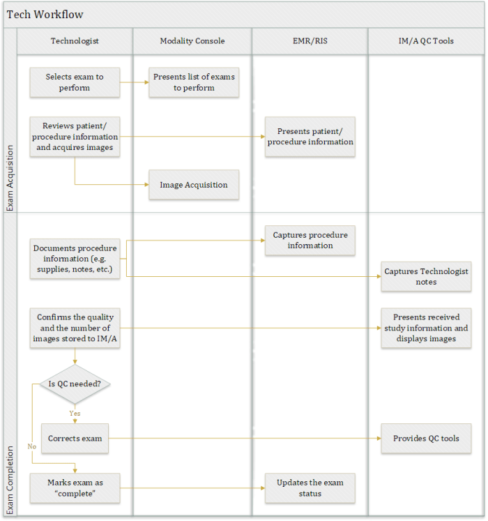
In the early days of Radiology, data entry errors by Radiology Technologists (aka Techs) were common. Their attention was on the patient and the operation of the modality, not the clerical task of typing in data, after all. To address this, something called a DICOM Modality Worklist (aka DMWL) was developed and adopted.

Essentially, this took the textual patient and imaging procedure order information entered into the HIS or RIS (i.e. the order placer), and sent it to some system as an HL7 ORM message (an order). The structured patient/order information was then provided to modalities using the DICOM protocol (because this is the language they speak). DMWL could be provided by the RIS or PACS or some form of broker system that spoke both HL7 and DICOM.

This allowed trained clerical workers (or physicians), combined with software that validated the data entered (where it could), to pass the information to the modality workstation where it could be mapped into DICOM objects, without having to ask Techs to enter this info. The productivity and information quality gains were significant.

It is worth noting that the order provides other value than just eliminating duplicate data entry. It represents a work instruction, and it is used in scheduling and billing. Where image acquisition is not scheduled or billed for, orders are typically not created.

Enter Enterprise Imaging

As we enter the era of Enterprise Imaging, there are lots of lessons that we can learn from the solved problems in areas like Radiology.

For example, when capturing a photo in a Wound Care clinic, it has to be associated with the correct patient (obviously), but there is likely other pertinent info that should be captured, such as the anatomical region imaged and any observations by the physician.

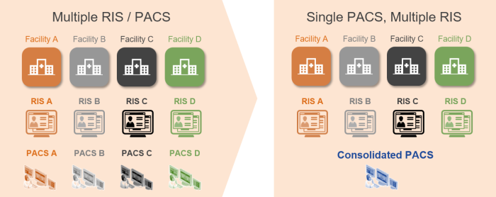
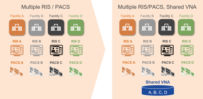
In Enterprise Imaging, orders are often not placed. In many areas, the imaging is often not the primary task, but one that used to support clinical work.

If orders are not placed, how can we at least provide the benefit of passing textual patient data to the image capture device or application to reliably associate patient (and perhaps encounter or procedure) data?

Even if orders are placed, most of the devices and applications used in Enterprise Imaging cannot accept an HL7 message and do not speak DICOM. Some form of broker would likely be required yet again.

Enterprise Information Interoperability for Enterprise Image Capture

One hope that we have is the adoption of the new HL7 FHIR standard. Based on REST-based API design methods, it is much easier to integrate with different devices (especially mobile devices) than HL7 v2.x messaging and DICOM interfaces are. Other methods used are to generate a URL from the EMR, with all the info provided in parameters, that launches the image capture application/device in context. Another method is to use HL7 messaging to populate the VNA database with patient, encounter and order/procedure information (essentially a copy of what the EMR has), and use a tool or API (perhaps the DICOMweb™ Query API, QIDO-RS) to query this system to get the necessary information.

Don’t Forget the Metadata and Supporting Information

This still leaves the issue of how to reliably and consistently capture the information that goes with the image(s)—notes, anatomy info, findings, technical exam info, observations, etc. In DICOM, when this type of information is needed, a SOP Class is defined. The header of the SOP Class object specifies where all this metadata should go. This is one of the primary principles of interoperability: a defined format and data scheme, with a clear and shared meaning.

Assuming that not all Enterprise Images will be generated in, or converted into, DICOM format, the definition of the metadata schema may be left to be defined by the implementing vendor.

In addition to the clinical and technical data, sooner or later, someone is going to be looking for operational data for use in analytics and process improvement, so it will need to be captured (on some level of detail), as well.
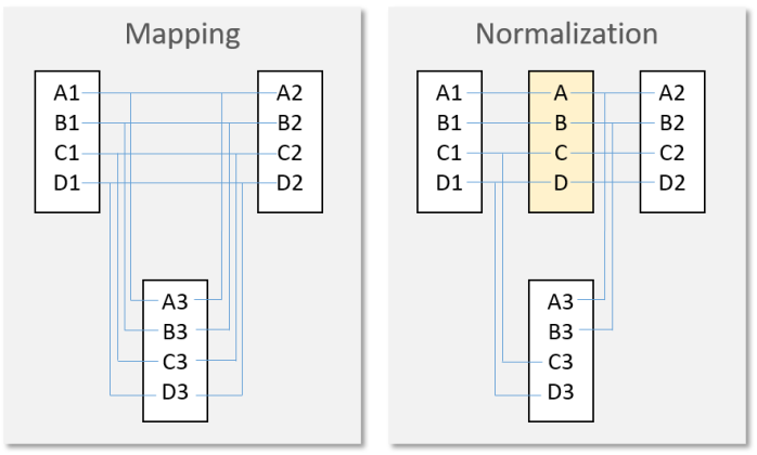
Consistent Terms

And, even when we have a common schema, if the terms used within the scheme are not consistent, we end up spending an enormous amount of time doing mappings or integrating terminology services (and even then, never fully addressing all cases).

To Acquire or Not to Acquire

If we think about Enterprise Imaging that is not “ordered”, what triggers the acquisition of an Enterprise Image? Is it up to the clinic or individual care provider to make the judgement? Should a published set of best practices define this? Would the EMR have logic, based on the patient’s condition or care pathway, to prompt or force the user to acquire and store the image(s)?

Enterprise Imaging Acquisition Protocols Needed?

If we consider the different ways that images can be captured (still, video), the subject in frame (cropping, zooming), lighting, etc., and the ability to capture a single image or a set of images, do we also need some form of a book of protocols to guide the person acquiring the images? Should certain images contain a ruler (or object of known size) to allow the image to be calibrated for measurements?

The Cost of Doing Nothing

If we consider the impact of not having methods to avoid data entry errors, or not having a common schema, not having common terms, and not even having a common communication protocol or best practices for acquisition workflows, what hope does Enterprise Imaging have?

Even with options for all these things, imaging and information devices are still struggling to be interoperable with departmental and enterprise applications, as described in this Healthcare IT News article, “Nurses blame interoperability woes for medical errors”.

The Future is Now(ish)

This is why the mission and output of the joint HIMSS-SIIM Enterprise Imaging workgroup (charter in PDF here) is so important. The space needs to be better defined, with acquisition workflow practices, data formats, schemas, terms, and protocols outlined.

If we simply try to copy what is done in Radiology into Enterprise Imaging, it will create too much of a burden on the people asked to capture these images, and they won’t do it, frankly. Unlike the reimbursement in Radiology, they often have little incentive to spend the extra time to capture, index and upload images to the EMR when they are focused on the patient.

But, if we ignore the benefits that come with the controls and methods we have developed and matured over the years in Radiology, we risk having to re-learn all the same lessons again. And that would be very sad (and expensive, and wasteful, and unsafe…).

Add on top of all this the increasing need to share this data across different enterprises for continuity of care and the importance of interoperable data portability/liquidity is critical.

The fundamental healthcare informatics knowledge and business analysis skills developed by imaging informatics professionals, through on-the-job experience and membership in educational/research societies like SIIM, will be important in determining the right mix of proven concepts that apply, and new methods and innovations. Without a supply of talent to foster the change, nothing will change.

In Conclusion…

When dealing with such an undefined space, people often relish the idea of “doing it right this time”. I would urge anyone involved in this space to reflect on what has been accomplished in mature fields like Radiology, as there are a lot of “right things” that we may be taking for granted. With a little modernization, we can still get continued value out of what we have already achieved.